Metatarsalgia pain at the ball of the foot
Home » Foot & Ankle Conditions – Consultant Podiatric Surgeon » Forefoot » Metatarsalgia pain at the ball of the foot
Metatarsalgia leads to pain or burning under the ball of the foot, often worsened by activity. Treatment works to reduce pressure, calm irritation and restore comfortable movement.
Arthritic Metatarsophalangeal Joint
Arthritis of the lesser metatarsophalangeal joints, the joints of the ball of the foot excluding the big toe joint, most commonly affects the second metatarsophalangeal joint.
Reasons behind the development of arthritis include trauma, healed Freiburg’s disease and inflammatory joint disease to include rheumatoid arthritis.
Non-surgical management can include:
- Steroid injections
- Alteration of footwear
- Activity modification
- Insoles
Surgical management can include:
- Removal of bony outgrowth around the joint and remodelling Joint replacement
- Removal of the base of the toe (avoided if possible).
There are several joint replacements available, the indications for each of these are influenced by a person’s age and activity levels. Joint replacements for this condition include the Cartiva implant.
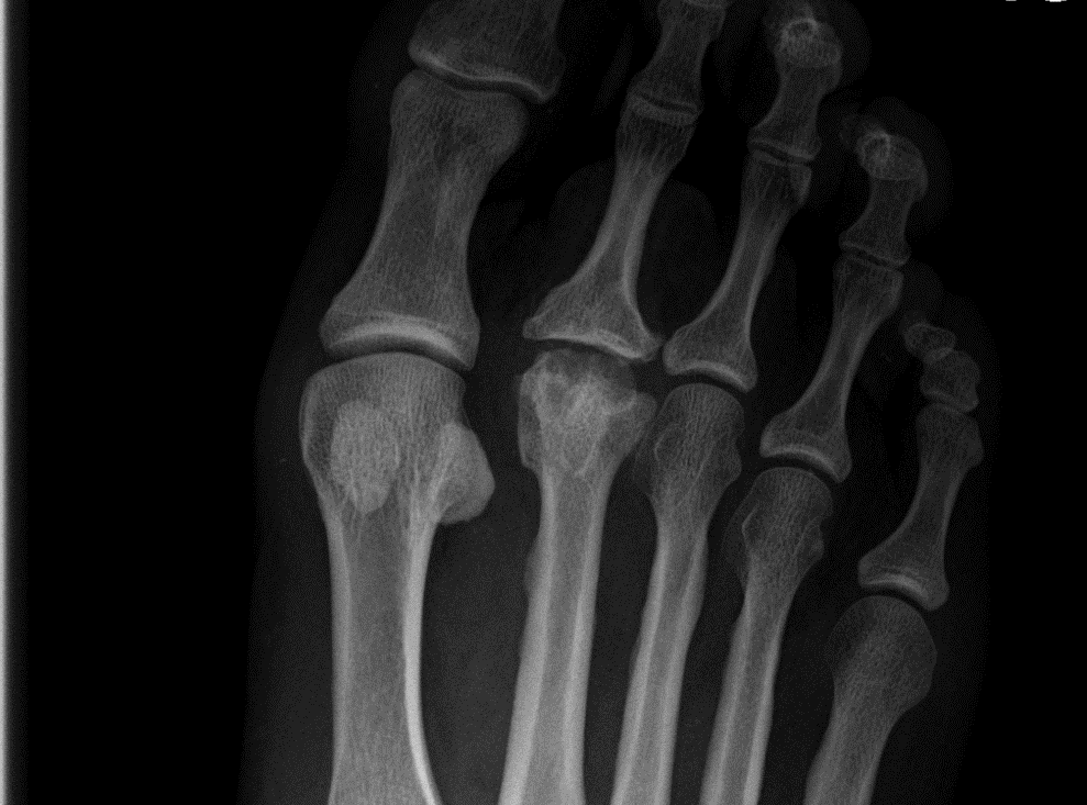
Arthritic second metatarsophalangeal joint with flattening of the metatarsal head and base of the toe bone (proximal phalanx), cyst formation and new bone formation on the sides of the metatarsal head. Overall the joint loses its normal appearance and appears more square.

Grossly arthritic second metatarsophalangeal joint associated with a large bunion and second toe deformity
Capsulitus
Capsulitis (cap-su-li-tis) is a common condition affecting the ball of the foot, the second metatarsophalangeal (meta-tar-so-fa-lan-ge-al) joint being the most commonly affected. This condition is often likened to walking around with a pebble in the shoe with the pain worsening when walking bare foot or when treading on the raised paving stones around traffic lights, going on tip toes or running. There are several different reasons as to why this problem occurs including overuse injuries, bunion deformities, long or prominent metatarsals and inflammatory conditions such as inflammatory arthritis i.e. rheumatoid arthritis.
The Plantar Plate
Initially the joint capsule and the fluid within becomes inflamed but as the condition progresses the joint capsule can become stretched or torn. The area of damage is related to a thickening of the capsule called the plantar plate. The plantar plate provides stability to the toe and prevents it from elevating and when damaged this restraining mechanism is lost and a hammer toe can develop. Often the condition is progressive and early management is essential to prevent worsening of the condition and the toe position.
Conservative and medical treatment includes:
- Alteration of activity
- Footwear modification
- Calf stretching (tightness in of the calf muscles can produce overload of the forefoot and increased pressure on the metatarsal heads and joint capsules)
- Insoles
- Steroid injections
Surgical management
Includes correcting any length abnormalities or prominence of the metatarsals and thereby reduce the force placed through the joint. This procedure is called a metatarsal osteotomy. Repair of the plantar plate. There are varying grades of plantar plate rupture and if the plate is not too damaged a repair may be possible. The video contains live surgery and shows the method of a plantar plate repair: Warning – Graphic Content
Tendon transfers are utilised if the plantar plate is beyond repair and the elevated position of the toe requires correction. The tendon transfer is performed at the base of the toe and involves relocating the tendon on the underside of the toe to the top so to pull the toe down into a corrected position. Correction of the hammer toe through either an arthoplasty or arthrodesis, to include any in or outward deviation of the toe.
Corn / Callus / Hard skin of the ball of the foot
Skin lesions at the ball of the foot are common and develop due to increased pressure when walking. The problem can either be isolated to one area, of the ball of the foot, or across the whole of the forefoot. In the case where there is an isolated lesion this is often related to a prominent metatarsal or due to redistribution of pressure secondary to the development of a bunion, tailor’s bunion and hallux limitus (stiff big toe joint). Hard skin formation across the whole of the forefoot can be related to certain foot types such as a high or low arched foot as well as tightness within the calf muscles.
Non-surgical treatment can include:
- stretching exercises
- insoles / padding
- footwear alteration
- Debridement / reduction of the corn
Surgery would address length or prominence issues by cutting and repositioning of the bones. This repositioning makes them less pronounced on the sole of the foot and thereby reduces the amount of pressure. Often hard skin can develop due to related problems including bunions or a tightness of the calf muscle. If stretching exercises have failed to resolve this the calf muscles can be lengthened through a procedure called a gastrocnemius recession.
Fractures
Metatarsal Fracture
Fractures of the foot can be caused by an acute injury such as sudden traumatic incident e.g. falling off of a kerb or through repetitive stress. Fractures that occur through repetitive stress and develop over time are termed stress fractures, pain is experienced when walking as can swelling, but is relieved by rest unlike an acute / traumatic fracture where displacement can occur and are commonly easily identifiable, stress fractures take time before they become apparent on x-ray, up to four weeks, and therefore an MRI is a more sensitive investigation as changes within the bone can be seen very early.
Management of stress fractures includes rest and a walker boot. If left untreated the pain and immobility can continue and may worsen with pain developing elsewhere in the foot trough an altered walking pattern. Stress fractures often maintain their alignment and unlike some acute / traumatic fractures do not require surgery.

Stress fracture of the midshaft of the second metatarsal (long bone of the foot). This stress fracture has developed due to overload from the short first metatarsal and an increase in the patient’s activity. The fourth and fifth metatarsals display a congenital bony bridge (synostosis)

Displaced midshaft fracture of the second metatarsal requiring surgical reduction due to displacement and elevation
Neuroma
A neuroma is a benign, non-cancerous condition where a nerve enlarges and produces pain. In the foot the most common place to develop a neuroma is at the ball of the foot between the metatarsal heads. Symptoms include burning, shooting and pins and needles (parathesia) into the associated toes. The pain is often relieved by removal of the shoe and rubbing the affected area.
The Most Common Neuroma
In the foot the most common neuroma is a Morton’s neuroma and is present between the third and fourth metatarsal heads (ball of the foot) and sends nerve pain into the third and fourth toes. Neuromas can develop between the adjacent metatarsal heads but a Morton’s neuroma is the most common.
Non-surgical treatment for a neuroma includes:
- Calf stretching exercises
- Footwear alteration
- Insoles
- Steroid injections
If non-surgical management fails to alleviate the pain surgery may be required.
Surgery includes either:
- Cutting the ligament above the neuroma to reduce pressure onto the nerve, much the same as a carpel tunnel release
- Excision of the nerve (neurectomy).
Depending on the size of the neuroma cutting of ligament above the neuroma may be indicated as this can be done through a minimally invasive technique (key hole surgery). A video of this technique can be viewed by clicking on the link below:
http://www.osteomed.com/SBO_Lower/KobyGard.aspx – (this link contains a video of live surgery – caution advised)
For more information about neuromas please click on the link below:
http://www.aofas.org/footcaremd/conditions/ailments-of-the-big-toe/Pages/Mortons-Neuroma.aspx
Osteochondrosis: Freiburg’s disease / Avascular Necrosis
Avascular Necrosis
Osteochondrosis (os-te-o-con-dro-sis) is a condition that affects bones that are still growing, the term avascular necrosis is used in an adult or if the bones have stopped growing. In the forefoot the second metatarsal is most commonly affected as this is the longest of the lesser metatarsals but is not exclusive to this bone. Both conditions involve an interruption to the blood supply to the bone which is often due to injury of a repetitive nature.
Freiburg’s disease
The term Freiburg’s disease is used when the condition affects the second metatarsal and most commonly occurs in females around the age of twelve. This condition produces pain during and especially after activity and may be associated with swelling and redness of the joint.
Management of this condition should be rapid as further trauma produces long term damage and arthritic changes of the joint. Within the first weeks of the condition developing x-rays often show nothing abnormal and MRI is the best investigation as this can diagnose the condition within the first few days of onset.
The condition is managed through immobilisation and activity modification. Over time and if allowed to rest the blood supply returns and if there has been alteration of the shape of the metatarsal a full recovery should be made. However, if the metatarsal has flattened and there is damage to the cartilage, over time, there is an increased chance of arthritic changes developing.
Tailor's Bunion
Tailor’s Bunion Deformity
A Tailor’s bunion is like a bunion deformity but occurs on the outside of the forefoot and involves the fifth metatarsal and little toe. The metatarsal moves towards the outside of the foot with the little toe moving towards the inside of the foot, the reverse of a bunion. The joint becomes prominent and rubs onto footwear and due to the poor alignment of the joint pain can also occur from the joint itself.
Tailor’s bunions can be managed by wearing a roomy shoe but if this fails to manage the problem surgery may be required. Surgery involves cutting and re-positioning the bone back into the foot. The bone is held together with screws.

Right foot view moderate sized bunion deformity and tailor’s bunion of the 5th metatarsal

Left foot showing correction of a bunion deformity and tailor’s bunion with screw fixation